Membranes:
A
membrane generally consists of two layers of tissue a layer of epithelium underlain and supported by a layer of connective tissue. (Some membranes consist only of thin layers of connective tissue, with secretory cells scattered through the matrix.) Since membranes contain epithelial tissue, theyre typically found lining body cavities, surrounding organs, and lining tubes that lead out of the body. The skin is also a membrane.
Mucous Membranes:
Mucous membranes, as youve probably guessed, contain epithelium (usually columnar epithelium) that is specialized to secrete mucus. Mucous membranes line tubes and organs that lead to the outside of the body, such as the digestive, respiratory and reproductive passages.
Mucus secreted by mucous membranes helps protect the digestive, reproductive, and respiratory tracts from infection by viruses and bacteria. In the digestive tract, mucus provides a protective barrier against digestive enzymes and acids that would damage the lining of the tract. Mucus membranes in the mouth and throat provide lubrication that protects against damage during chewing and swallowing. Mucous membranes in the female reproductive tract provide lubrication that protects against damage during sexual intercourse and during childbirth.
Serous Membranes:
Serous membranes, of course, secrete serous fluid. These membranes are found lining the internal organs and the body cavities, and they consist of a layer of simple squamous epithelium underlain by a thin layer of connective tissue. These membranes secrete serous fluid that cushions and lubricates the internal organs. In addition, they help support the internal organs and hold them in position; they also compartmentalize body cavities, which helps to slow infections.
Within the thoracic and abdominal cavities, most internal organs are surrounded by two layers of serous membranes. The cavity within which an organ sits is lined by a
parietal serous membrane, and the organ itself is surrounded by a
visceral serous membrane. For example, the heart sits within a cavity called the
pericardial cavity. The pericardial cavity is lined by the
parietal pericardial membrane (
parietal pericardium), and the heart itself is surrounded by the
visceral pericardial membrane (
visceral pericardium).
The parietal and visceral membranes secrete serous fluid that fills the space between them, lubricating and cushioning the organ(s) contained within the cavity.
Pleural Membranes:
Each lung is contained within a
pleural cavity. Naturally, each pleural cavity is lined by a parietal pleural membrane and the lung itself is surrounded by a visceral pleural membrane. Uniquely, the pressure of the serous fluid secreted by these membranes is slightly
less than is normal air pressure. This means that the air inside the lungs presses outward with more force than the serous fluid surrounding the lungs presses inward. That pressure difference is what keeps the lungs inflated. If the pleural cavity is punctured and air enters the space between the parietal and visceral membranes, its pressure can force the lung to collapse. This is known as a
pneumothorax, and needless to say, is a very bad thing.
Pericardial Membranes:
The heart, as mentioned a moment ago, sits within the
pericardium, and is surrounded by parietal and visceral pericardial membranes. Were it not for the serous fluid secreted by these membranes, and for the layer of fat surrounding the pericardial cavity, the heart would surely be damaged as it beat because of abrasion against the ribs and sternum.
Peritoneal Membranes:
Peritoneal membranes (
peritoneum) line the abdominopelvic cavity and most of the organs lying within it. Smooth muscles of the stomach and intestines cause these organs to move somewhat during the digestive process. Were it not for lubrication from the serous fluid secreted by parietal and visceral peritoneal membranes, these organs might be damaged by abrasion.
Synovial Membranes:
Synovial membranes line joint cavities where two bones come together to form a
movable joint. (Where two bones come together is a
joint or
articulation. Not all joints are movable movable joints are known as
synovial joints.) Unlike most membranes, synovial membranes contain no epithelium. Synovial membranes secrete
synovial fluid into the space between two bones, which lubricates the ends of the bones and prevents them from damaging each other as they rub together. If inadequate amounts of synovial fluid are produced, bones may abrade each other as they move; splinters of bone in the joint cavity may cause irritation and swelling of surrounding tissues. This inflammation of joint tissues is known as
arthritis.
So far, no one has developed a synthetic lubricant thats as slippery as is synovial fluid.
Meninges:
Meninges (
meningeal membranes), like synovial membranes, contain only connective tissue. These membranes surround the organs of the
dorsal cavity that is, the
brain and the
spinal cord.
There are three layers of tissue in the meninges: the
pia mater, the
arachnoid mater, and the
dura mater. The pia mater (the name means tender mother) lies closest to the brain and the spinal cord. Blood capillaries within the pia mater supply the brain and spinal cord with oxygen and nutrients. The arachnoid mater is named for its spiderweb-like appearance. The space between the arachnoid mater and the pia mater (the
subarachnoid space) is filled with
cerebrospinal fluid, which cushions and lubricates the brain and spinal cord. The dura mater (hard mother) is a tough and relatively inflexible outer membrane that surrounds and protects the central nervous system. It fuses to the underside of the skull and to the inside of the
neural canal in vertebrae. The dura mater contains larger blood vessels that split into the capillaries of the pia mater.
A bacterial or viral infection of the meninges can cause inflammation and swelling of these membranes. This is
meningitis, and the increased pressure can cause damage to the delicate underlying tissues of the brain or spinal cord.
A
subdural hematoma occurs when veins bridging the dura mater and arachnoid mater are torn. This is typically a result of head trauma, such as may occur during an automobile accident, for instance. Blood leaks into the space between the the dura mater and arachnoid mater, and the increased pressure can cause severe brain damage.
An
epidural hematoma is similar to a subdural hematoma, except that its caused by tearing of arteries in the dura mater. This causes blood to fill the space between the dura mater and the skull. As in a subdural hematoma, the increased pressure can quickly cause severe brain damage.
The Cutaneous Membrane:
The
cutaneous membrane is the skin. It consists of an outer layer of stratified squamous epithelium underlain by a thicker layer of loose connective tissue and/or adipose tissue. Well discuss the cutaneous membrane in some detail in the next chapter.
Organs and Organ Systems:
A tissue, as you recall, consists of cells of the same general type that perform a common function. An
organ is a structure made of two or more tissue types that together perform a common function.
For example, the stomach contains an inner layer of epithelium that is supported by connective tissue. Layers of smooth muscle surround the epithelial and connective tissue layers. Nervous tissue relays messages from the central nervous system to the muscles and glandular tissue of the stomach.
Groups of organs that work together to perform a common function make up an
organ system. For example, the stomach is part of the
digestive system. For the sake of convenience, the various organ systems are often divided into various categories, according to what they do.
Organ systems that are important in
support and movement of the body include the
integumentary system, the
skeletal system, and the
muscular system.
The
nervous system and the
endocrine system allow the
integration and coordination of body functions.
Transport of substances throughout the body and
immunity against disease are the responsibility of the
cardiovascular system and of the
lymphatic system.
Absorption of nutrients and oxygen and
excretion of metabolic wastes is the responsibility of the
digestive system, the
respiratory system, and the
urinary system.
Finally, the
reproductive system is responsible for
well
reproduction.
In the pictures below, you can see many of the components of the various organ systems and their positions in the body. The remaining chapters of this series will focus on the individual organ systems. Well start with the one organ system you can observe without taking a scalpel to your acquaintances the
Integumentary System.
 Superficial Body Structures
Here you can see the skin (the cutaneous membrane), the mammary glands,
and some of the major superficial muscles.
 Superficial and Deep Muscles
Here you can see many of the superficial muscles (mostly on the subject's right) as well as deeper muscles (mostly on the subject's left). Some of the more superficial veins and arteries can be seen as well. The femoral nerve can be seen extending into the leg.
 The Viscera
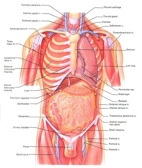
Here, most of the overlying muscles have been removed, revealing the underlying viscera. The ribcage surrounds and protects the heart and lungs, as well as most of the stomach and liver. The diaphragm is a muscle that separates the thoracic cavity and the abdominopelvic cavity. The mesentery is a layer of tissue that hangs down over the intestines like an apron; it secretes fluid that lubricates the intestines.
 The Internal Organs
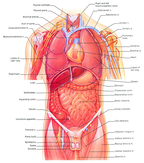
The ribcage and mesentery have been removed, exposing most of the organs of the thoracic and abdominopelvic cavities. Most of the respiratory system can be seen, as can most of the organs of the digestive system. Most of the major veins and arteries can be seen. Most of the male reproductive system is visible.
 Deep Body Structures
The internal structure of the heart and lungs is visible. The ureters, which transport urine from the kidneys to the urinary bladder are visible. Most of the female reproductive system (which lies much deeper than does the male reproductive system) is visible. Some of the deeper veins and arteries are visible. The deep muscles of the upper leg are visible.
 Deeper Body Structures
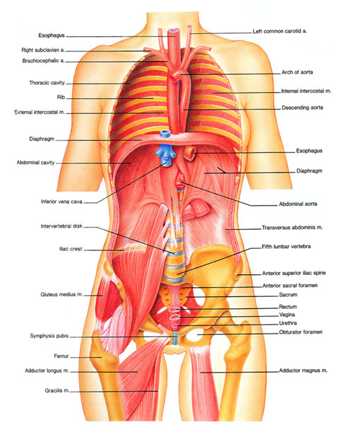
The esophagus can be seen, lying deep to the trachea. The kidneys can be seen, lying behind the main portion of the abdominopelvic cavity. Most of the major deep blood vessels can be seen. More of the deep muscles of the leg can be seen.
 Really Deep Body Structures
Here you can see the back of the ribcage. You can see how the esophagus, the vena cava (the body's major vein) and the aorta lie almost as far back as do the vertebrae. Many of the bones that form the framework of the body are visible.


 Linear Mode


